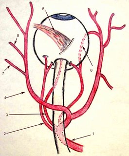
What is the blood supply and drainage for the choroid?

The blood supply to the choroid comes ultimately from the ophthalmic artery (#1 in Figure). There are variations but quite posterior branches that will become the central retinal artery (#3 in Figure), and ciliary arteries on each side of the optic nerve. These vessels divide 2 long posterior ciliary arteries and about ~20 short posterior ciliary arteries that enter the eye immediately adjacent and around the optic nerve. The short posterior ciliary arteries directly supply the choroid and the long posterior ciliary arteries travel in the suprachoroidal space anteriorly (#6 in Figure) then supply the choroid anteriorly via recurrent branches. The ophthalmic artery (#4 in Figure) continues to provide branches for the posterior (#7 in Figure) and anterior (#8 in Figure) ethmoidal vessels. The superior oblique muscle is shown for orientation ( #9 in Figure).
Blood in the choroid circulates through the choriocapillaries and larger vessels of the choroid to drain into the 4-6 vortex veins (#1 in the figure). These
 emerge just posterior to the equator in quadrants (#2 in the figure). The superotemporal and superonasal vortex veins will drain into the superior ophthalmic vein. The inferonasal and inferotemporal (#3 in the figure) vortex veins will drain into the inferior ophthalmic vein. These vessels will eventually exit via the cavernous sinus (# 5 in the figure).
The superotemporal ophthalmic vein usually exits the eye directly adjacent to or underneath the superior oblique tendon. This has clinical implications for approaches to surgery in which the superior oblique tendon is recessed.


0 Comments:
Post a Comment
<< Home