What are the paranasal sinuses?
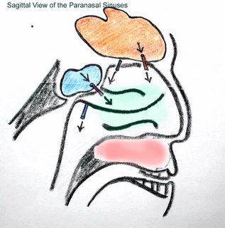
 The paraorbital sinuses include the frontal sinus above the orbit (orange), the ethmoid sinuses (green) in the medial wall of the orbit, the sphenoid sinus (blue) that is posteriorly located and the maxillary sinus (purple-pink).
Below, the sinuses are shown in a sagittal view. The maxillary sinus (red-pink) and the ethmoid sinuses would not be visible in the midline plane that shows the drainage from the sinuses (arrows). It is evident that the maxillary sinus is close to the teeth. As such pain from the sinus may be referred to teeth. This occurs in maxillary sinus infections. Occasionally normal teeth have been removed from patients with life threatening fungal sinus infections because this source of referred pain was unrecognized.


1 Comments:
Hi, my name is jorge huaman m.d. and I head the resident´s trainning program at my hospital in Lima Peru (Hospital Militar Central). I found an amazing tool for teaching and learning eye anatomy.
congatulations
jorge huaman m.d.
Post a Comment
<< Home