
Optic Canal, Optic Chiasm
The opening to the optic canal is the optic foramen at the apex of the orbit in the less wing of the sphenoid bone. From its location at the junction of the roof and medial wall of the orbit the optic canal is directed medially, upward moving posteriorly to the middle cranial fossa. This direction is useful to distinguish the optic canal from the superior orbital fissure on CT scan. The optic canal is on the same horizontal plane as the upper portion of the superior orbital fissure so they will often be seen in sections of the CT scan together. The optic canal houses the optic nerve, ophthalmic artery and sympathetic fibers from the cartoid plexus. A line drawn from the top of the nose to the auditory canal will approximately form the inferior border of the optic foramen. The dura of the optic nerve merges with the periosteum as the nerve enters the optic canal. The optic canal measures about 12 mm in length and about 7 mm in width but there are quite a few variations (Akdemir et al).
Upon exiting the optic canal, the optic nerve now lies medial to the internal carotid artery as seen in the picture below.
 Identify the structures in the photograph: arrow 1. optic nerve
2. superior oblique muscle transected and removed 3. superior rectus muscle transected and removed
4. ophthalmic artery
5. posterior ciliary arteries
6. posterior ethmoidal artery
7. anterior ethmoidal artery
8. supraorbital artery
9. lacrimal gland
10. ophthamic-internal carotid
As th optic nerve exits the orbit, it lies above the ophthalmic arteries but below the anterior cerebral arteries and anterior communicating arteries, which join to complete the anterior circle of Willis (see figure). The optic nerve passes over the cavernous sinus extending medially to join the fellow optic nerve and form the optic chiasm. The optic nerve and chiasm is therefore medial and superior to the cavernous sinuses. The optic chiasm lies anterior to the pituitary gland stalk (see figure link). These anatomic relationships become important when discussing the effect of aneurysms and tumors on the visual pathway.
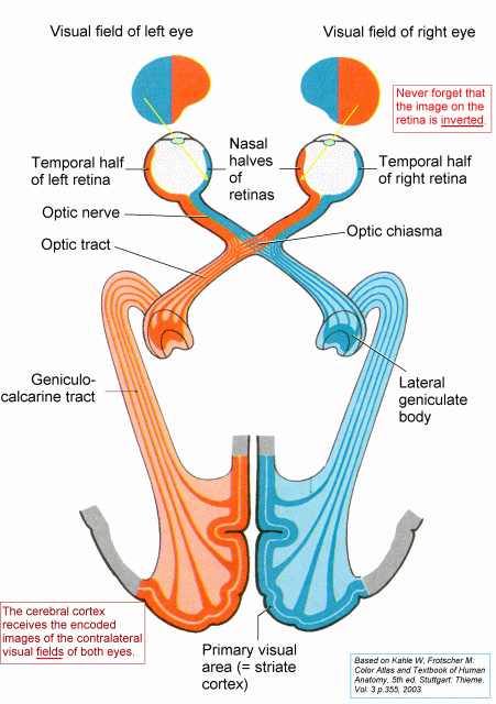
The general and simplistic projection of visual fields in relation to the organization of optic nerve fibers as nasal fibers (temporal fields) cross through the optic chiasm. Therefore the right visual field is projected to the left side of the brain (see link). However, there are some important details of these anatomic relationships that the ophthalmologist needs to remember:
1. extramacular nasal and inferior retinal fibers (superotemporal fields) cross in the anterior portion of the chiasm (Wilbrand's knee red fiber in Figure). Hence the relationship to pituitary tumors.

2. Nasal macular fibers cross in the posterior part of the chiasm.
3. Temporal fibers remain uncrossed.
4. Macular projections are located centrally in the nerve and chiasm and account for most of the fibers!!!


0 Comments:
Post a Comment
<< Home