
Lacrimal Gland Histology
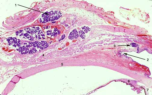
Photomicrograph (above) shows a low power image of an exenteration specimen that has the eye intact with the adnexal structures. The lobules of the orbital portion of the lacrimal gland (1) are near the orbital septum but lie under the levator muscle (4). The fornix of the upper eyelid (2) lies immediately adjacent to the accessory lacrimal gland of Krause(3). The glands of Krause are accessory lacrimal glands having the same structure as the main gland. They are placed deeply in the subconjunctival connective tissue (mainly) of the upper fornix between the tarsus and the inferior lacrimal gland, of which they are offshoots. There are some 42 in the upper and 6 to 8 in the lower fornix. They are thus found largely on the lateral side. Their ducts unite into a rather long duct or sinus which opens into the fornix. Similar glands are found in the caruncle. The Glands of Wolfring or Ciaccio are also accessory lacrimal glands, but larger than the glands of Krause. There are 2 to 5 in the upper lid situated actually in the upper border of the tarsus about its middle between the extremities of the tarsal glands or just above the tarsus. The excretory duct is large and short and lined by a basal layer of cubical cells and a superficial layer of cylindrical cells like the conjunctiva on which it opens. The sclera (5), ciliary body(6) and iris (7) are all identifiable.
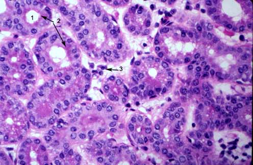
Photomicrograph (above) shows a higher magnfication of a human lacrimal gland complete with acinar structures that contain lumens (1) and protein rich acinar cells that secrete lysozyme, tear lipocalin, lactoferrin and IgA. The reddish granules are secretory vesicles replete with protein. Some lumens are filled with prtotein that is being secreted. Lymphocytes and plasma cells are scattered in the interstitium.
The lacrimal gland situated above and lateral to the eye in the orbit secretes the tears and into ducts in upper fornix. The lacrimal gland and its tears exist in animals which live in air. Fish do not have lacrimal gland. The lacrimal gland consists of an orbital or superior portion; and a small palpebral or inferior portion; which are continuous. The orbital portion is lodged in its fossa on the anterior and lateral part of the roof of the orbit. It is shaped like an almond.
The lacrimal gland consists of a lobules and is a tubulo-racemose gland with short branched gland tubules somewhat similar to the parotid. The acini consist of two layers of cells placed on a thin hyaline basement membrane and surrounding a central lumen. The basal cells are myoepithelial in character while the acinar cells are cylindrical, and secrete fluid into a series of ducts of increasing size until becoming the excretory duct.


0 Comments:
Post a Comment
<< Home