VITREOUS
 click on photo for a larger image
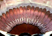
Above -dark pigmentation on the anterior border of the vitreous base (VB). The serrated border of the retina is the ora serrata. Measurements of the ora serrata vary and include a range from 5.75-7.5 mm from the limbus nasally and about 6.5-8.5 mm temporally. In my experience, drawn from transillumination of autopsy and surgical eyes the rectus muscle tendons insert within the vitreous base, but vary with respect to the position of the ora serrata. The medial rectus tendon inserts well anterior to the ora serrata. The studies by White et al. drawn from only 10 post mortem eyes vary from what is published by Wolf and vary again from what is shown in the academy BCSC series. A large study of autopsy eyes taking into consideration the size of the eyes would clarify this issue.
Below- transillumination highlights the pigmentation of the vitreous base as it straddles the ora serrata.

The vitreous has distinct attachments to ocular structures. It is attached anteriorly in a circumferential band extending from the posterior pars plana to a few millimeters behind the ora serrata in what has been termed the vitreous base. Traction exerted by the vitreous body at the base results in hyperpigmentation of the underlying pigment epithelium and is evident grossly. The vitreous is also attached to the retina over retinal blood vessels and at the optic disc. These attachments are important to understanding vitreous traction, retinal tears, and retinal detachment, for which vitrectomies are sometimes performed.
[Previous Page] [Next Page]
[Table of Contents] [Text on Mission for Vision]


0 Comments:
Post a Comment
<< Home