
CONJUNCTIVA
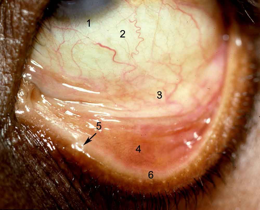
The conjunctiva covers the posterior surface of the eyelids (palpebral conjunctiva), curves anteriorly at the fornix to reflect onto the anterior surface of the eye as the bulbar conjunctiva. There are subtle histologic differences in the conjunctiva of the lid margins, tarsus, fornix, and bulbar conjunctivae. The conjunctiva covering the lid margin and bulbar conjunctiva is a modified nonkeratinized, stratified squamous epithelium. The tarsal and fornix conjunctiva is covered by stratified cuboidal to columnar epithelium of varying thickness. This epithelium is unusual because it retains some squamoid features, such as numerous desmosomes, yet has a microvillus surface architecture. Goblet cells are abundant over the tarsus, fornix, and specialized areas such as the plica semilunaris. Goblet cells are scarce near the lid margin and adjacent to the cornea at the limbus. Test your ability to identify the various areas of the conjunctiva by identifying the type of conjunctiva and its histologic characteristics in the pictures below. 

Click on the photo to enlarge and identify the structures that are numbered. Click here to link to the answers.

The histology of the caruncle can be seen in the link below on Mission for Vision.
[Previous Page] [Next Page]
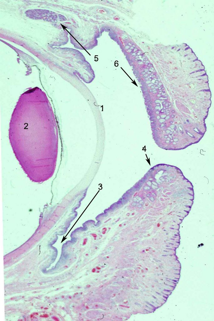
Click on the photo to enlarge and identify the structures that are numbered. Click here to link to the answers.

The histology of the caruncle can be seen in the link below on Mission for Vision.
[Previous Page] [Next Page]


0 Comments:
Post a Comment
<< Home